2013 Student Projects
Back to REU homepage
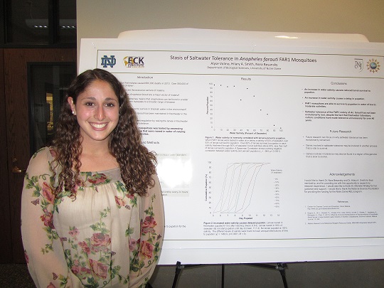
Stasis of Saltwater Tolerance in Anopheles farauti FAR1 Mosquitoes
Alyse Volino, Hilary A. Smith, Nora Besansky
 Alyse Volino
Saltwater tolerance in anophelines is a trait with important implications in disease transmission and human health. The existence of saltwater tolerant anopheline species means that anophelines can be found in a wider geographic distribution, which translates to a broader range of disease transmission. Mosquitoes of the species Anopheles farauti are a major vector of malaria. Anopheles farauti has been found to survive in brackish water in the environment, but a colony of the FAR1 strain of An. farauti has been maintained in freshwater in the laboratory since 1967. Removing selection pressure for osmoregulation by raising the larvae in freshwater may have resulted in loss of salinity tolerance. Thus saltwater tolerance of FAR1 mosquitoes was tested by assessing survivorship to pupation of larvae that were reared in water of varying salinities. Water salinity was found to be related to the percentage of larvae that survived to pupation, as well as the amount of time it took larvae to develop to pupation. As water salinity increased, survivorship to pupation decreased. Treatment with water at salinities above 60% that of seawater proved to be detrimental to pupation: no more than 40% of larvae in any treatment higher than 60% of seawater survived to pupation. Also, time to pupation increased as salinity increased, from 7 days in larvae reared in freshwater up to 11 days in larvae reared in 90% of seawater. The ability of FAR1 mosquitoes to survive to pupation in water of low to moderate salinities shows that the saltwater tolerance of Anopheles farauti has not been lost, even though freshwater laboratory culture conditions have made tolerance unnecessary for over 40 years.
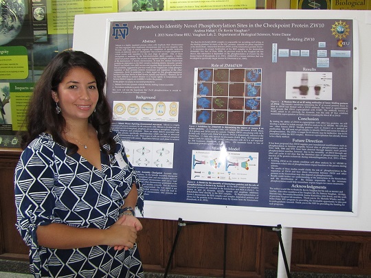
Approaches to Identify Novel Phosphorylation Sites in the Checkpoint Protein ZW10
Andrea Hakaj, Kevin T. Vaughan
 Andrea Hakaj
Mitosis is a highly regulated process in which cells duplicate their chromosomes and then segregate these duplicated chromosomes into two new daughter cells. Because each stage of mitosis requires the completion of specific molecular events, an elaborate series of “checkpoints” has evolved to couple completion of critical events with transition to the next stage of mitosis. One of these checkpoints is mediated by the Rod-Zw10-Zwilch (RZZ) complex, a highly-conserved protein complex that recruits and regulates multiple essential proteins at the kinetochores of mitotic chromosomes. At least five distinct kinetochore proteins are dependent on the RZZ complex, including dynein, dynactin, spindly, Mad1/2 and BubR1. How RZZ recruits and regulates these proteins is not known. One possibility, suggested by previous work in the Vaughan lab, is that phospho-regulation of RZZ proteins controls recruitment and regulation of the other components. Zw10, in particular, is a likely target for this phospho-regulation because it has been shown to bind dynein, spindly and Mad1/2. However, Zw10 has been difficult to analyze because it is bound tightly to kinetochores and because limited reagents are available to isolate the protein. In this project, we propose to use novel protocols to: 1) release Zw10 into cytosolic extracts thereby making it more accessible, and 2) to evaluate methods to purify this released Zw10. Building on previous work in the Vaughan lab that identifies Zwint-1 as a novel substrate for AurB (Kasuboski et al., 2011), we will treat HeLa cells with ZM447439 to block recruitment of the RZZ complex to kinetochores thereby enhancing the cytoplasmic pool of Zw10. After evaluating the impact of ZM treatment, we will test immunoprecipitation with anti-Zw10 antibodies and purification with recombinant zwint-1 as methods to isolate Zw10. Using which ever method provides biochemical quantities of Zw10, we will subject purified Zw10 to MS/MS analysis to identify phosphorylation sites. These sites will be evaluated using mutagenesis and transfection assays. Together this work will test the hypothesis that Zw10 phosphorylation is crucial to regulation of progression through mitosis.
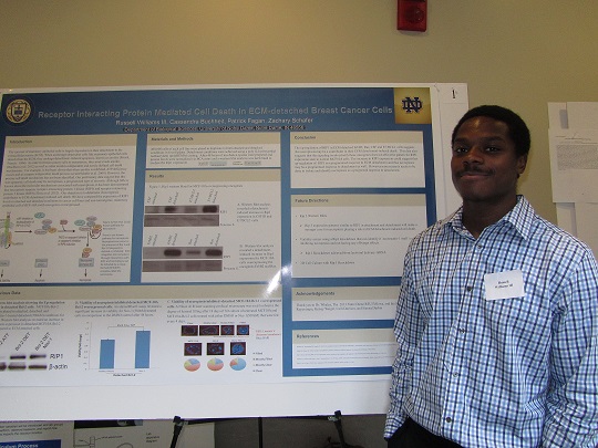
Receptor Interacting Protein Mediated Cell Death in ECM-detached Breast Cancer Cells
Russell Williams III, Cassie Buchheit, Patrick Fagan, Zachary Schafer
 Russell Williams III
The survival of mammary epithelial cells is largely dependent on their attachment to the Extracellular-matrix (ECM). When anchorage-dependent cells, like mammary epithelial cells, detach from the ECM, they undergo detachment-induced apoptosis, known as anoikis (Frisch, Francis. 1994). In order for breast cancer cells to metastasize, they must evade anoikis (Buchheit et al. 2012) as well as other anoikis-independent and poorly defined cell death mechanisms. For example, it has been shown that when anoikis is inhibited, ATP deficiency occurs and cells still die (Schafer et al. 2009). However, the precise cell death mechanism has not been identified. Our preliminary data suggest that this non-apoptotic cell death may be necroptosis, a programmed type of necrosis. Although little is known about the molecular mechanisms associated with necroptosis, it has been demonstrated that necroptosis requires receptor interacting protein 1 kinase (RIP1) and receptor interacting protein 3 kinase (RIP3) (Zhou et al. 2012). Our objective is to determine if necroptosis contributes to ECM-detachment induced cell death. We have
compared the expression of RIP1 and RIP3 levels in attached and detached conditions in cancer cell lines and non-tumorigenic, mammary epithelial cells (MCF-10A) with oncogenes overexpressed. We found that in MCF-10A cells with Bcl-2 overexpression, RIP1 is up-regulated in ECM-detached conditions. Additionally, our lab has found that in the absence of apoptosis in Bcl-2 overexpressing cells the viability of cells treated with the necroptosis inhibitor Nec-1, increases compared to Bcl-2 cells treated with DMSO. This result suggests that these cells undergo necroptosis in the absence of apoptosis when detached from the ECM. We hypothesize that cancer cells may prevent the increase of Rip1 levels blocking necroptosis from occurring in ECM-detached conditions. Understanding ECM-detachment induced cell death processes is paramount for finding targets for novel therapeutic treatment approaches that target metastatic cancer cells
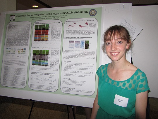
Interkinetic Nuclear Migration in the Regenerating Zebrafish Retina
Rebecca Marton, Manuela Lahne, David Hyde
 Rebecca Marton
Zebrafish can regenerate lost photoreceptors due to Müller glial cells that reside in the inner nuclear layer of the retina. Müller glia divide in response to light-induced photoreceptor damage to produce neuronal progenitor cells that ultimately differentiate into new photoreceptors. During eye development, neuronal progenitor cells follow a pattern of movement referred to as interkinetic nuclear migration (IKNM). In IKNM, neuronal progenitor cell nuclei migrate from the basal region of the retina, where DNA is replicated, to the apical surface, where they undergo cell division. Recently, the Hyde lab demonstrated a similar Müller glial nuclear migration in the adult zebrafish eye during the regeneration response. While studies implicated actin/myosin interactions as a driving force of IKNM in development, the mechanisms that govern Müller glia nuclear migration during regeneration are not yet understood. Immunohistochemistry experiments that I conducted using phalloidin labeling revealed the presence of filamentous actin in processes in the trailing side of Müller glial nuclei during the regeneration response, suggesting the involvement of actin/myosin interactions in the regenerating retina. To assess the role of actin/myosin interactions in IKNM, I am identifying targets to inhibit actin filament formation. One such class of targets are α-actinins, cross-linking proteins that stabilize actin filaments. Quantitative real time PCR (qRT-PCR) experiments were used to determine what zebrafish α-actinin subforms change in expression during retinal regeneration. Upregulation of α-actinin1 and α-actinin3a beginning prior to the onset of Müller glial nuclear migration suggest that these subforms may be involved in IKNM, while downregulation of α-actinin3b and α-actinin2 suggests that these subforms may be associated with dying photoreceptors. In situ hybridization experiments confirmed α-actinin1 expression in Müller glia at the onset of IKNM but not in the undamaged retina. Together, qRT PCR and in situ experiments provide strong evidence for α-actinin1 as a target to inhibit actin filament formation in Müller glia during IKNM.
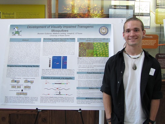
Development of Visually Impaired Transgenic Mosqitoes
Brandon Gutierrez, Matthew Leming, Joseph E. O'Tousa
 Brandon Gutierrez
Aedes aegypti is the primary vector for transmission of yellow fever and dengue fever. Treatment of these diseases is costly and not effective, causing vector control to become a promising method for disease prevention. The visual systems of mosquitoes likely provides sensory input driving behavioral responses. This study is designed to further understand the role of visual systems in vector behaviors such as blood-feeding, oviposition, flying, and mating. Rhodopsins are a protein family responsible for initiation of the phototransduction cascade. The role of a major rhodopsin, Aaop1, as well as vision as a whole in vector behavior will be evaluated by developing visually impaired mosquitoes. Two transgenic strategies are being utilized to create these animals. The first strategy is to develop a transgenic line of mosquito in which Aaop1 will be knocked out through the use of transcription activator-like effector nucleases (TALENs). The second transgenic approach is targeted ablation of photoreceptor cells by using the Aaop1 promoter to drive retinal expression of michelob_x, a cell death gene. A construct containing the promoter-driven death gene will then be inserted into the genome of Ae. aegypti through utilization of a transgenic line containing a recombination site, or through use of the piggyBac method to insert the construct into the genome at a random location. The construct will be flanked by a gypsy insulator sequence which will serve to prevent any repression caused by the localization of the insert.Upon successful generation of these transgenic lines, behavioral assays will be used to determine the effect of visual impairment on vector behaviors using wild type mosquitoes as a control group.
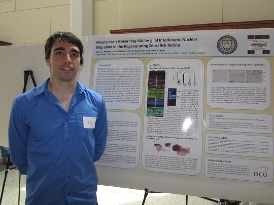
Mechanisms Governing Müller glial Interkinetic Nuclear Migration in the Regenerating Zebrafish Retina
Ciarán J. Mooney, Manuela Lahne, Kristin Ackerman, David R. Hyde
 Ciarán J. Mooney
Inherited retinal diseases, such as retinal macular degenerations, result in the irreversible loss of photoreceptor cells leading to blindness. Unlike humans and other mammals, lower vertebrates, such as zebrafish, have the ability to undergo retinal regeneration following injury. Thus, zebrafish are a popular model organism to study retinal regeneration. Following light-induced injury in the zebrafish retina, Müller glial cells undergo dedifferentiation, enter the S phase of the cell cycle and produce neural progenitor cells that ultimately replace the lost or damaged photoreceptor cells [1]. During this process, Müller glia undergo interkinetic nuclear migration (IKNM), where their nuclei migrate along a basal-apical axis in phase with cell cycle progression [2]. While IKNM has been well characterized during neuroepithelia development, our laboratory has only recently observed IKNM in the adult zebrafish during retinal regeneration. Preliminary data found that actin filaments accumulate at the basal side of Müller glial nuclei that migrated apically during retinal regeneration. Additionally, Müller glial IKNM in regenerating adult zebrafish retinas decreased following inhibition of rho kinases, which phosphorylate and activate the myosin light chain, and facilitate actin-myosin mediated contraction [Unpublished observation]. Moreover, a microarray study carried out in our lab found the non-muscle myosin heavy chain myh10 is upregulated in the zebrafish retina following injury [3]. As such, I am investigating the role of the actin cytoskeleton and myh10 during Müller glial IKNM in the regenerating zebrafish retina.
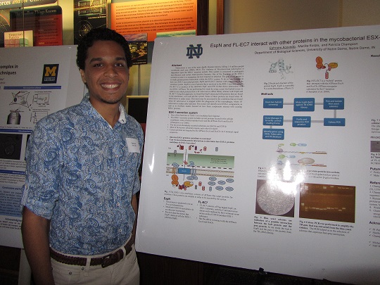
EspN and FL-EC7 interact with other proteins in the mycobacterial ESX-1 secretion system
Ephraim Acevedo, Marika Kuspa, Patricia Champion
 Ephraim Acevedo
Tuberculosis is one of the most deadly diseases known, killing 1.4 million people worldwide each year (Who, 2012). The virulence of Mycobacterium tuberculosis is dependent upon the ESX-1 secretion system. This system is conserved in pathogenic mycobacteria and certain Gram-positive bacteria. One of the functions of the ESX-1 secretion system is to manipulate the host response to infection. The goal of my project is to study protein-protein interactions within the ESX-1 system. To do so I am focusing on how two ESX-1 associated proteins,EspN and FL-EC7, interact with components of the secretion system. EspN is a novel protein that is involved in the ESX-1 secretion system. FL-EC7 is a construct of the substrates EspC and CFP-10 that shows interactions with two ESX-1 ATPases. We are performing this study by using a yeast two-hybrid screen in order to see which proteins from a M. tuberculosis cDNA library interact with EspN and FL-EC7. Using this approach, we found that EspN interacted with proteins that are found in the membrane, cell wall and the cytosol. Some of these proteins are found when M. tuberculosis is under stress. This stress may be associated to the conditions that are found when M. tuberculosis is trapped within the phagosome of the macrophages, where M. tuberculosis resides after infection. This screen will identify novel ESX-1 components, as well as to facilitate studies focused on how these components interact with existing ESX-1 proteins.
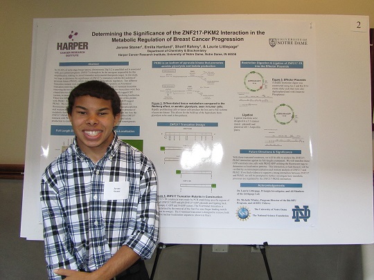
Determining the Significance of the ZNF217-PKM2 Interaction in the Metabolic Regulation of Breast Cancer Progression
Jerone Stoner, Emilia Hartland, Sharif Rahmy, Laurie Littlepage
 Jerone Stoner
In 20-30% of early-stage breast tumors, chromosome 20q13.2 is amplified and is associated with poor patient prognosis. ZNF217 is thought to be the oncogene responsible for this amplification, making it a novel biomarker and potential therapeutic target. In this study, we hope to determine the significance of ZNF217’s interaction with the M2 isoform of pyruvate kinase (PKM2), which plays a vital role in metabolic regulation. Two different truncation mutants were made from the full-length ZNF217 protein: an N-terminal truncation removing the first five zinc-finger binding motifs and a C-terminal truncation removing the two putative nuclear localization sequences. These two truncations were then cloned into two different vectors, N-terminally-tagged and C-terminally tagged GFP vectors, to ensure that there was no tag interference in ZNF217 function during localization. These four different ZNF217 mutants will be used to study the interaction of this protein with PKM2. Each construct will be transfected into 293 cells along with RFP-tagged PKM2. The localization patterns of ZNF217 and PKM2 will then be monitored via fluorescence microscopy. The interaction will also be verified by co-immunoprecipitation and western analysis of ZNF217 and PKM2. Future experiments testing the presence of metabolic factors in cells overexpressing ZNF217 will provide more insight into how the ZNF217-PKM2 interaction may affect metabolic regulation. It is expected that ZNF217 interacts with PKM2 to regulate metabolic processes like glucose intake and lactate production to create hypoxic and low-nutrient environments that promote tumorigenesis.
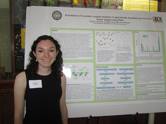
Knockdown of G-protein coupled receptors in adult female Anopheles gambiae and Aedes aegypti using RNAi
Katelyn Carothers, Douglas Shoue, Mary Ann McDowell
 Katelyn Carothers
Malaria, yellow fever, and other insect transmitted diseases are a growing worldwide concern, responsible for millions of human deaths every year. Increased human population and urbanization of tropical areas contribute to the continued spread of the diseases, as well as growing insect resistance to current insecticides and concerns of their effect on the environment and on human health. G-protein coupled receptors, already a common drug target in humans, contribute to a variety of physiological processes in eukaryotes, and are under consideration as novel targets for insecticides. The targets of this study included AgOAR45B, an octopamine receptor, and AgNPF10626 and AgNPF2733, the respective receptor and ligand of neuropeptide F in in Anopheles gambiae. We attempted to knock down the production of AgOAR45B in adult female Anopheles gambiae via infection with a recombinant Sindbis virus containing the antisense sequence to the AgOAR45B, in order to induce RNA interference in the cells. We also transcribed dsRNA for AgNPF10626 and AgNPF2733 for direct injection into the mosquito as an alternative to the viral pathway of gene knockdown. We then quantified the expression of the genes in infected and control mosquitoes using qRT-PCR. Knockdown of gene expression by RNAi can contribute to greater knowledge of the target gene’s role in the organism. Future studies may include screening of agonists and antagonists for AaOAR45B and AaNPF10626, and their development as components in future insecticides.
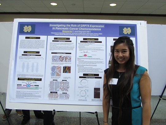
The Role of GRP78 Expression in Pancreatic Cancer Chemoresistance
Shayna Hu, Reginald Hill
 Shayna Hu
Pancreatic ductal adenocarcinoma (PDAC) is the fourth leading cause of cancer related deaths in the United States with a 5 year survival rate of 6%. A major contributing factor for the high mortality of PDAC is the chemoresistant nature of the disease. GRP78 is an endoplasmic reticulum (ER) chaperone protein that protects cells from cell death and promotes cell survival. It is highly expressed in many tumor types and may play a significant role in tumor progression and chemoresistance. We found high GRP78 expression in pancreatic tumors from both human patients and a mouse model of PDAC, and this projects aims to investigate GRP78’s effect on chemoresistance. We plan to address this question by first overexpressing GRP78 in pancreatic cell lines to investigate its downstream targets. Next, we will examine the effect of inhibition on these targets on cell survival and chemoresistance. We have begun constructing vectors to overexpress GRP78 in stably transfected cell lines and in an inducible manner. We ultimately plan to develop a therapeutic strategy that targets the GRP78 pathway to inhibit tumor progression and increase susceptibility to currently available chemotherapeutics. Overall, this will allow us to significantly increase the survival rate of the people impacted by this disease.
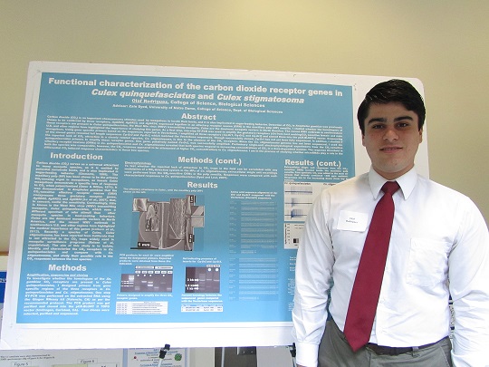
Functional characterization of the carbon dioxide receptor genes in Culex quinquefasciatus and Culex stigmatosoma
Olaf Rodriguez
 Olaf Rodriguez
Carbon dioxide (CO2) is an important chemosensory stimulus used by mosquitoes to locate their hosts, and it is also implicated in sugar-feeding behavior. Detection of CO2 in Anopheles gambiae was previously shown to be conferred by three receptors, AgGR22, AgGR23, and AgGR24, expressed together in an olfactory receptor neuron (ORN) in the maxillary palp (MP) sensilla. I studied whether the homologues of these receptors are present in Culex quinquefasciatus, the West Nile virus (WNV) transmitting mosquito. Culex are the dominant mosquito vectors in North America. The recent WNV outbreak in southwestern U.S. and other regions have highlighted the importance of studying this genus. As a first step, one-step RT-PCR was used to amplify the gustatory receptors (Gr) from host seeking/CO2 sensitive female and male mosquitoes. Using gene specific primers based on the sequences reported in Vectorbase, I amplified all three receptors (Cq-Gr1, Cq-Gr2, and Cq-Gr3) and cloned them into the pCR-BLUNT-II vector. Sequencing of the cloned genes revealed full length sequences Cq-Gr2 and Cq-Gr3, which matched the Vectorbase sequences. Though successfully cloned, Cq-Gr1 has not yet been fully sequenced. In addition, I studied if the reported lack of CO2 attraction in a closely related species, Cx. stigmatosoma, is due to the absence of the CO2 receptors. Since the Cx. stigmatosoma genome has not been sequenced, I used Cx. quinquefasciatus primers to amplify these receptor genes. One gene, putatively named Cs-Gr2, was successfully amplified. Preliminary single-unit electrophysiological experiments from the CO2-sensitive olfactory receptor neurons (ORNs) in Cx. quinquefasciatus and Cx. stigmatosoma revealed that both species respond to increasing concentrations of CO2 in a dose-dependent manner. The response threshold in both the species was comparable, however, the CO2 response appeared to be attenuated at higher CO2 concentrations in Cx. stigmatosoma. I am in the process of studying whether microRNAs play a role in the differential CO2 induced response characteristics in these two species.
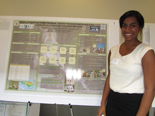
Population Genetic Structure of Balinese Long-Tailed Macaques (Macaca fascicularis)
Julia Resil, Amy Klegarth, Hope Hollocher
 Julia Resil
Increases in human population and subsequent urban development have a marked impact on the environment. Animal habitation in the vicinity of humans has a prospective possibility of having their dispersal patterns and species gene flow experiencing extreme distortion. This study examines genetic variation and population structuring of Balinese long-tailed macaques (Macaca fascicularis) in varying degrees of anthropogenic influence. Long-tailed macaques are not only known for their dispersal abilities but are also known extensively throughout Southeast Asia; conversely Balinese macaques are honored at Hindu temple sites and are offered varying degrees of food provisioning. This research is designed to examine the influence of varying levels of provisioning of macaque genetic variation. In spite of Balinese macaque’s large population sizes, my hypothesis is that food provisioning will result in decreased dispersal, ultimately decreasing gene flow and more importantly decreasing genetic variation. This study is designed to determine the degree of genetic variation on Bali based on correlation of genetic distance and geographical distance. This method of study has been previously used to examine gene flow on Singapore via mitochondrial and nuclear datasets. A previous incomplete dataset from Bali generated a haplotype network with limited success on genetic variation relation to population structure. By using mitochondrial DNA to make haplotype networks it will be possible to visually analyze differences and similarities associated with Singapore and Bali respectively. Further tests on genetic variation will include more analysis with mitochondrial on the number of social groups per site and the number of matrilines per group. Furthermore we will be able to achieve sharper data on genetic variation that was previously discovered by a study that used nuclear DNA from Bali macaques. Future goals of this on going project would be to give preliminary data to be used for macaque management and study of anthropogenic environmental influences on other mammalians and eventually to other types of organisms.