
Medical Imaging Technologies
 Within one year of the 1895 discovery of X-rays by Wilhelm Röntgen,
images of the stomach and other organs were being used to identify and
locate tumors. Within 10 years hospitals around the world were using
the new imaging technology to help diagnose and treat patients. Innovations
in medical imaging have consistently focused on capturing as much information
as possible … as accurately and as quickly as possible.
Within one year of the 1895 discovery of X-rays by Wilhelm Röntgen,
images of the stomach and other organs were being used to identify and
locate tumors. Within 10 years hospitals around the world were using
the new imaging technology to help diagnose and treat patients. Innovations
in medical imaging have consistently focused on capturing as much information
as possible … as accurately and as quickly as possible.
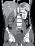
The emphasis in the last five years, according to Ken D. Sauer, associate professor of electrical engineering and director of the Engineering Honors Program, has been to lower the radiation a patient encounters during a scan. “Unfortunately,” says Sauer, “this results in lower-quality images, unless we do something to aid the reconstruction process.”
Much of Sauer’s research focuses on improving the methods for image reconstruction in computed axial tomography (CAT or CT) and magnetic resonance imaging (MRI) scans. “We don’t work on the machine itself or design the X-ray emitters,” he says. “We take the data that’s created during the scan and apply very specific algorithms (part of a software package) to help us better reconstruct the images. When these algorithms are successful, they are built into special-purpose hardware, which increases the speed and accuracy of image reconstruction.”
 Medical
imaging technologies involved in radiation therapies present different
challenges. For example, researchers at Notre Dame, in conjunction with
researchers at the University of Iowa and the University of Maryland
School of Medicine, are addressing some of these image issues. Danny
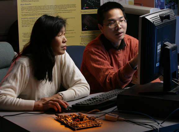
Z. Chen, professor of computer science and engineering, has
been working with Milan Sonka, professor of electrical
and computer engineering at the University of Iowa, on a National Institutes
of Health project to refine image segmentation for organ contouring. “Medical
imaging is a physician’s eye into the body,” says Chen. “Even
when you know the disease, you have to know exactly where it is located
to avoid damaging healthy tissue and vital organs. You need all of this
information before you can accomplish effective treatment.”
Medical
imaging technologies involved in radiation therapies present different
challenges. For example, researchers at Notre Dame, in conjunction with
researchers at the University of Iowa and the University of Maryland
School of Medicine, are addressing some of these image issues. Danny
Z. Chen, professor of computer science and engineering, has
been working with Milan Sonka, professor of electrical
and computer engineering at the University of Iowa, on a National Institutes
of Health project to refine image segmentation for organ contouring. “Medical
imaging is a physician’s eye into the body,” says Chen. “Even
when you know the disease, you have to know exactly where it is located
to avoid damaging healthy tissue and vital organs. You need all of this
information before you can accomplish effective treatment.”
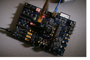 Chen and X. Sharon Hu, associate professor of computer science and engineering and director of the department’s graduate studies program, are also developing hardware-based methods to enhance image registration. One of the major problems in medical imaging is that scans of the same area in the same body do not always match. “This is very important,” says Hu, “especially for mapping organs that move (change shape, such as the heart and lungs), tracking the amount of radiation absorbed by a tumor, or assessing the growth or reduction of a tumor.” Whether for radiation treatment or computer-aided surgery, fast and accurate image registration is vital.
Chen and the Notre Dame computational medicine group have also developed new algorithms and software called SLS, which are based on graph theory models and computational geometry techniques. These are helping to produce intensity-modulated radiation therapy plans that reduce the treatment time by more than 60 percent while providing higher quality images. The technology is currently being used in the University of Maryland Medical Center and the Helen P. Denit Cancer Center in Montgomery General Hospital in Olney, Md., where it is aiding in the treatment of patients.
For more information on medical imaging technologies at Notre Dame, visit http://www.nd.edu/~engineer/publications/signatures/2007/medimaging/index.html