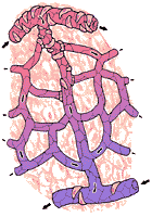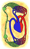
fig 240. Structures of the Capillary
Unit IV - Circulatory System
Chapter 13
Capillaries and Lymphatics
 |
fig 240. Structures of the Capillary |
1. Function and Structure of Capillaries:
The flow of blood to a particular area depends on the state of its arterioles. The exchange of material between the blood so delivered and the cells occurs through the capillaries. The tissues continuously produce carbon dioxide and other products of activity; they also use oxygen and food stuff continuously. The supply of oxygen and food depends on the movement of these substances across the capillary wall, from the blood to the tissue. The removal of carbon dioxide and waste depends on movement in the reverse direction.
The capillaries, where these exchanges occur, are thin tubes whose construction is such as to make these exchanges very easy. See Figure 240. Unlike the other blood vessels, the capillaries consist only of the endothelium, which is the same as the lining layer of the arteries and arterioles. A typical capillary is 1 mm long. Its diameter is about 8 - 10 micra (1 micron = 0.001 mm). The thickness of the wall across which the exchanges of materials occurs is about 0.2 micra. The total surface area of the capillaries in the resting body is about 40 square meters.
 |
Although the blood usually remains in the capillaries for only a second, their walls are so thin that there is plenty of time for the necessary exchanges to occur. The great number of capillaries further insures adequate exchange. There are probably about 50, 000, 000, 000 capillaries; ordinarily only 2, 000, 000, 000 are employed at any time in normal exchange. The remaining capillaries are sealed off from the circulation by muscles which surround their arteriolar end. These muscles, called the pre-capillary sphincters, open and close at intervals. By rotation of the precapillary sphincters which are open, (Figure 241) the capillaries "in service" are continuously changed, and all tissue areas are brought intermittently into functional contact with the blood. In certain organs, such as the brain, heart, and kidney all the capillaries are open at all times. In muscular exercise, the increased requirements of the muscle are met by the opening of most of the precapillary sphincters of the active muscle.
2. Colloid Osmotic Pressure and Transcapillary Fluid Movements:
The capillary walls are so thin that the blood pressure is enough to drive almost all the water in the blood across them. This does not actually happen, for the pressure of the blood is compensated by the fact that the basement membranes do not permit the passage of the dissolved proteins of the blood. The way in which the proteins prevent the complete removal of water is described in the next paragraph. When pressure is applied to a liquid, the molecules of the liquid gain energy. They become more mobile, and they tend to move away from the area where the pressure was applied. If some of the molecules in the liquid cannot move away (if, for example, the liquid is being forced through a membrane through which these molecules cannot pass), some of the pressure energy which could have been used to move the movable molecules is wasted; each of them will move less easily than if the entire pressure energy were available to move the movable molecules. These molecules will still move, but not as well as if the "non-passing" molecules were not present.
If a pressure is applied on the other side of the membrane, where only the easily movable molecules are present, its effects will be fully realized, as a movement across the membrane to the low pressure side. When equal pressures are applied to a liquid on both sides of a membrane, but the liquid on one side contains molecules which cannot pass through the membrane, the overall effect will be net transfer of liquid from the side that does not contain the "non-passing" molecules to the side that does.
The movement of liquid to the side with the "non-passing" molecules can be prevented by increasing the pressure on that side. The amount of pressure required to prevent this movement is called the osmotic pressure exerted by the non-passing molecules. This value can be considered just as if it were a true fluid pressure.
In the capillaries, the non-passing molecules are the proteins. The osmotic pressure which they exert is normally 26 mm Hg, that is to say, it would take 26 mm Hg pressure to cause water to leave the capillary. Proteins in the interstitium exert a minor osmotic pressure of about 1 mm Hg which pulls in the opposite direction, so the net pressure required to extravasate water from the capillary is actually closer to 25 mm Hg.
At the beginning of the capillary, the fluid pressure is about 35 mm Hg. Since this is opposed by the osmotic pressure of the proteins inside the capillary (26 mm Hg) and assisted by the osmotic pressure of the proteins in the interstitial fluid (1 mm Hg), there is a net pressure of 10 mm Hg available to force water out of the capillary and into the spaces of the tissue.
At the end of the capillary, the fluid pressure has fallen to about 26 mm Hg. Now no pressure is available to force fluid out of the capillary since the protein osmotic pressure is greater than the fluid pressure. In fact, the reverse occurs. There is a net pressure tending to restore fluid to the capillaries amounting to 9 mm Hg.
 |
 |
 |
Thus, the overall balance of forces at the arteriolar end of the capillary tends to cause fluids to leave. At the venular end, fluids are restored. The forces are approximately (though not quite) equal, and the amount of fluid in the tissue space remains relatively constant almost all the time. The relationships described above were first suspected by Starling who gave a very clear account of them before 1900. They are illustrated in Figure 242.
3. Lymphatic System:
The balance of forces within the capillary is usually very slightly in favor of forcing fluid out of the capillary. If this were not compensated, it would result in continual loss of fluid into the tissue spaces. This probably does, in fact, occur. The lost fluid (lymph) is, however, picked up in a system which is called the lymphatic system, and returned by it to the blood.
The lymphatic system has its roots in the lymphatic capillaries of Figure 243. These are very much like the blood capillaries in their distribution, but they differ from them in being blind ended. The fluid filtered from the capillaries in excess of that which is taken back into them passes into the tissue spaces and is picked up in the lymphatic capillaries. These fuse, making larger and larger vessels (lymph vessels). The larger lymph vessels are equipped with valves, which are so arranged that the fluid cannot move backward to the lymph capillaries, but only toward the main lymph channels, which return the fluid to the heart. (Figure 244).
In the course of its passage through the lymphatic system, the lymph passes through a number of specialized structures, the lymph node. (Figure 244). These nodes, sometimes called lymph glands, produce lymphocytes (Chapter 9). In addition, the lymph glands function to remove from the lymph returning to the heart the foreign materials and bacteria which may have been picked up by the lymph in its passage through the tissue spaces. The lymphatic system is shown in Figure 245.
4. Disorders of the Capillary and Lymphatic System:
The lymphatics serve to take care of the fluid which leaves the capillaries which is in excess of what returns to them. Usually, this is a very small amount. In some conditions, where excessive amounts of fluid are formed, or where the return of fluid to the capillary is impaired, or if the lymphatics are blocked, fluid accumulates in the tissue spaces in large amounts. This accumulation is called edema.
Edema often develops in normal people, particularly in the feet and ankles when they have been sitting still for a long time. The reason for this is that the fluid pressure in these capillaries tends to be much higher than in other capillaries, because they are so far below the heart. The high fluid pressure is not balanced by the protein osmotic pressure and fluid therefore leaves the capillary. Movement of the legs ordinarily compensates for the increased fluid pressure because this movement aids in returning blood from the veins past valves which prevent the full fluid pressure from being exerted on the capillaries. When the legs are not moved, as for example, in the case of passengers in a long car or airplane trip, the feet and ankles become edematous. This type of edema disappears rapidly when the feet are elevated above heart level, or as a result of muscular activity.
When the valves of the veins of the legs do not function well, the fluid pressure in the lower parts of the body is raised, whether or not there is motion. This occurs in varicose veins. These superficial veins of the legs stretch because of the fluid pressure; and thus become visible at the surface. In addition, the raised fluid pressure produces a chronic edema of the ankles.
The reason that this or any other type of edema does not result in continuous fluid loss has to do with the fact that as fluid is lost, the pressure of the fluid outside the capillaries builds up. The swollen tissues become tight and eventually the increased pressure outside the capillaries compensates for the increased pressure inside them. In treating varicose veins the same thing can be accomplished, without as much tissue swelling, by the use of elastic stockings to supply extra pressure which keeps the blood fluids from entering the tissue spaces in excessive amounts.
Earlier it was noted that the heart could meet an increased work load by being stretched with blood to a larger volume. This is associated with increased intraventricular pressure during diastole. The veins, which must empty into the heart also show an increase in pressure, and in the same way, the fluid pressure of the capillaries increases. This leads to edema in many parts of the body. Of course, this edema does not usually develop in the normal person, since the normal heart rarely employs this mechanism in adjusting its work. A person with a damaged heart, however, is likely to use this mechanism, even at very low work loads. It is quite characteristic of right heart failure to see edema of the lowest parts of the body (dependent edema), even when activity is low. In left heart failure the left heart is enlarged; its pressure during diastole is elevated and the veins which empty into the left side of the heart (the pulmonary veins) show pressure increases. Sometimes this shows itself simply as difficulty in breathing. Sometimes edema develops in the lungs; this will be considered in more detail in Chapter ?.
The most dramatic types of edema are seen in children. In starvation, the formation of plasma proteins is reduced and therefore the protein osmotic pressure may not be sufficient to keep fluids inside the blood stream. Edema results. Usually, the edema fluid tends to accumulate in the abdomen rather than in the legs. This brings about a picture of a child with spindly legs and arms (because of starvation) and a swollen belly.
A kidney disease usually seen only in children is lipoid nephrosis. In this condition, the kidneys leak plasma proteins into the urine. The level of plasma proteins becomes low, and the fluid forces in the capillaries become unbalanced. Such children may become edematous quite suddenly. For reasons which are not well understood, their edema occurs in all parts of the body. A temporary relief of this edema is obtained by giving plasma protein concentrates by vein.
A form of edema which is quite familiar is the formation of a blister after a burn. Through the reflex, the arterioles of the burned area are dilated and the capillary fluid pressure rises. Besides this, the capillary walls in a burned area tend to become more permeable and pass proteins. 1The combination of increased fluid pressure and the loss of protein osmotic pressure (the proteins are ineffective in maintaining osmotic pressure unless they are confined by a membrane) results in the rapid loss of fluid in the burned area. As was noted before in the section on varicose veins, this does not continue indefinitely because the pressure of fluid in the blister balances the unbalanced forces in the capillary. If, on the other hand, the blister breaks, the counter pressure is lost and fluid continues to leave the capillaries in the affected area. This results in the oozing of the surface of a burned area.
When the burned area is small, this may not be a serious matter. When it is large, the oozing may occur so rapidly as to produce serious losses of plasma and circulatory collapse. This may be treated by giving plasma, and by the large scale application of counter pressure. For example, when a large area of the leg is burned, some surgeons find it helpful to enclose the leg in a plaster cast.
When bacteria are picked up in a lymph node, they are sometimes killed; sometimes they multiply in the node. The node responds to this by growing larger. This often results in a swelling of the node which can be felt through the skin, or seen directly. Swelling of lymph glands in the back of the neck is commonly seen when there are scalp infections. Swelling of the tonsils, which are lymph glands, occurs after certain infections of the throat.
Obviously, by removing bacteria, the lymph glands form a part of the defense system of the body. However, when the bacteria multiply in the lymph glands, they may act as a place from which bacteria may be spread. The original bacterial infection may have been a minor one; when the lymph gland is invaded, it may serve as a new focus of infection, from which bacteria may spread--either to other lymph glands or eventually back to the blood stream and the organs. This process is called metastasis, and is also important in determining the spread of some cancers.
Thus, for example, in cancer, some cancer cells may find their way into the lymph channels. When they reach the lymph glands, they may be stopped; but they will multiply there. Eventually they may fill up the lymph gland and move by way of lymph vessels to the next lymph gland. When the involved organ is removed, it is not enough to remove it alone. The cancer cells in the lymph nodes must also be removed. This calls for removing nodes, sometimes quite far away from the original cancer. In cancer of the breast, the lymph nodes which are often invaded are in the armpit. These should be removed if there is any suspicion of metastasis. In cancer of the tissues of the face, the entire system of lymph nodes of the neck must often be removed.
Continue to Chapter 14.