
A National Science Foundation Research Experiences for Undergraduates Site
Department of Anthropology, University of Notre Dame, Notre Dame, IN 46556

Guest Scientist
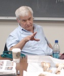
Prof. Donald Ortner of the Smithsonian Institution’s Museum of Natural History will be the guest scientist for the 2007 Program. He will work with each group to fine tune their research design as well as provide instruction on various aspects of paleopathology.
Emily Schach [North Carolina State] & Mary DeAgustino [University of Notre Dame]
This project will examine the tibial plateau (the articular surface on the proximal end of the tibia) for degenerative changes associated with destruction of the cartilage at the knee joint. You will score for bony growths (osteophytes), bone-on-bone contact (eburnation), porosity, and lipping of the margins. A group in last year’s osteology course collected data on degenerative joint disease of the distal femur, such that you could compare incidence among the patella with incidence of the patellar articular surface of the femur.

Ki Won Kim [UC-Berkeley] & Matthew Gasperetti [University of Notre Dame]
Parry fractures are a result of trauma to the mid to distal end of the ulna. Their name comes from “parrying” a blow – they are usually the result of holding your arm in front of your face to protect yourself. Colle’s fractures are a result of trauma to the distal end of the radius. They tend to be represent accidental falls wherein the individual places their arms straight out in front of their body as they fall to avoid hitting his/her head. You will be examining both the distal radii and ulnae.
Sarah Henkle [Tulane University] & Laura Major [Dickinson College]
Schmorl’s nodes are depressions on the superior & inferior vertebral bodies due tof weakening of the cartilaginous intervertebral disc. You will look for these indicators on the superior and inferior surfaces of lumbar vertebrae. Your challenge will be in identifying superior and inferior surfaces of the body (when only the body is present - which you will be able to do with practice), and in your statistical analysis. There are 5 lumbar vertebrae, but we will most likely not know which “number” most of the vertebrae are.
Stephanie Kalchik [University of Notre Dame] & Rachael Byrd [Fort Lewis College]
You will be examining the mandibular condyle for evidence of degenerative joint disease at the temporomandibular joint. You will examine porosity and bony exostoses on the mandibular condyle as an indicator of joint disfunction. While a complete project would focus on both sides of the joint (the mandibular condyle and the mandibular fossa of the temporal), you will concentrate on the mandible this summer. This project could certainly be continued to include the temporal in the future. The complex etiology will prove challenging in your analysis, but you should have a large sample size, and there is definitely degenerative joint disease in the group. In addition, there is a large amount of clinical literature on this topic.
Daniel Bradley [University of Notre Dame] & Alissa Jordan [Indiana University-Bloomington]
Osteomas are benign lesions, or harmless bony growths. They tend to appear on frontals and parietals, and are found almost exclusively on the skull. They are fairly common in archaeological populations, and you should be able to find a decent amount of comparative data. Although their etiology is not certain, this project will provide another facet of comparison for the Bab edh-Dhra’ group to other groups.
.


Previous NSF-REU Summers
A summary of previous research projects can be found: