|
ITS 2004 Summer Visiting Fellows Program
August
13, 2004 - ITS Mini-symposia on Bio-imaging at Argonne National
Laboratory
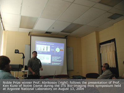
On August 13, 2004 the Institute hosted three speakers
at Argonne National Laboratory: Prof. Gabor
Forgacs from the University of Missouri-Columbia, Prof.
Masaru Ken Kuno from the University of Notre Dame, and
Prof. Rusty Lansford from the Beckman
Institute, California Institute of Technology. The mini-symposia
was of large interest, among the audience were 2003 Physics
Noble Laureat Prof. Alexei Abricosov and other distinguished
Argonne scientists.
ITS SYMPOSIA ON BIO-IMAGING
ARGONNE NATIONAL LABORATORY,
August 13, 2004
Useful biomechanics from the subcellular
to the organismal levels
Prof. Gabor Forgacs ~ University of Missouri
- Columbia
Abstract: I will overview our lab's activity
in the field of biomechanics with emphasis on those aspects
that are biologically useful. At the subcellular level, biomechanics
is used to improve on the yield of embryo cryopreservation.
At the cellular scale, biomechanics is employed to quantify
and thus better understand how white blood cells or tumor
cells may initiate exiting the circulatory system, respectively,
during inflammatory response and metastasis. At the supercellular
or tissue level we rely on biomechanics to optimize our efforts
in studies on morphogenetic processes, in particular “organ
printing”, an evolving technology to construct 3D functional
organ modules of desired shape using methods of rapid prototyping.
In these endeavors we use magnetic tweezers, atomic force
microscopy-based force micromanipulators, specifically designed
bioprinters and other special purpose, usually in-house built
instrumentation.

Dynamic
Visualization of Embryogenesis 
Prof. Rusty Langsford ~ Beckman Institute,
California Institute of Technology
Abstract: My research aims to dynamically
visualize and characterize avian embryonic development at
sub-cellular resolution. My primary focus is determining how
the brain and heart form and develop. Instead of labeling
and following small numbers of cells at a time, I am trying
to optically record all or nearly all the cells within a developing
embryo simultaneously. I will place fluorescent tags within
all avian embryonic cells using GFP-expressing retroviruses.
Cell and tissue movements in the developing embryos are recorded
using multispectral, time-lapse fluorescent microscopy in
3D. The recorded data is then analyzed using computers running
sophisticated cell-tracking and color discrimination software
capable of distinguishing the subtle movements that thousands
of individual cells make and identifying a handful of genes
that these cells express. The gene expression and cell migration
data collected using laser microscopes is then integrated
within MRI collected datasets in order to understand the complex
informational interactions that are occurring during development
within the spatial and temporal context of the maturing embryo.
Solution-based straight and branched
semiconductor nanowires
Prof. Masaru K. Kuno ~ University of Notre
Dame
Abstract: Long standing interest in understanding
and ultimately controlling crystal growth has recently materialized
as studies into new routes for making high quality metal and
semiconductor nanocrystals (NC), nanorods (NRs), nanowires
(NWs), as well as other higher order nanostructures. The discovery
that metal NCs have catalytic properties for promoting asymmetric
crystal growth has motivated studies into making 1D semiconductor
NWs. Recent investigations have led to the development of
synthetic techniques that include variations of traditional
vapor-liquid-solid growth, wherein chemically synthesized
or laser ablated metal NCs are used as catalyst particles.
Such routes take advantage of advances in NC syntheses to
overcome intrinsic droplet size limitations, ultimately allowing
one to create previously unattainable, narrow diameter NWs.
Other approaches include complete solution phase analogues
of VLS growth such as solution-liquid-solid (SLS) growth and
supercritical-fluid-solid (SFLS) growth.
Here the solution phase synthesis of narrow diameter (<
10 nm) straight and branched CdSe NWs is described. Crystalline
NWs with lengths between 1-10 microns are obtained using a
seeded solution approach, whereby NW growth is initiated using
Au/Bi core/shell NCs. Such wires may exhibit unique quantum
confinement effects given that the bulk exciton Bohr radius
of CdSe is 5.6 nm. Manipulating the reaction conditions allows
one to transition from straight to branched nanowires yielding,
tripod, v-shaped, and y-shaped NWs. Further variations in
the preparation lead to higher order NWs that exhibit multiple
branching points. In all cases, the presence of surface binding
surfactants yields soluble straight and branched NWs opening
up intriguing opportunities for future surface modifications
and/or surface functionalization chemistries. Such branched
wires also provide the distinct possibility of studying not
only size dependent optical and electrical properties of NWs
but also their shape dependent properties as well.
|